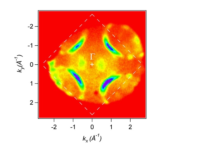
| 95 eV photon energy:
|
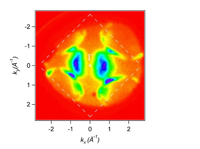
122 eV photon energy: |
|
|
|
The PEEM can not only be used to display magnified images of the sample
surface. When the projection lenses are set to display the focal
plane of the objective lens, the angular distribution of the emitted electrons
is obtained. In connection with an imaging
energy filter this can be used to display two-dimensional photoelectron
angular distribution patterns on the screen of the PEEM. In this
way spatial, angular, and energy resolution can be obtained simultaneously
within the same instrument. |
slice through Cu
Fermi surface ("dog bone"):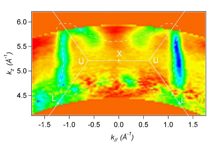
|
The figures show such angular distribution patterns of photoelectrons from the Fermi energy of a Cu(001) single crystal surface. Different photon energies yield different cuts through the three dimensional Fermi surface of Cu (pink = high, red = low intensity). The complete three-dimensional Fermi surface can be obtained rather quickly by acquiring a series of such images for different photon energies. A slice through a data-set of 59 images is shown in the bottom image. The well-known “dog-bone” shape of the Cu Fermi surface is clearly seen. Publication: Review of Scientific Instruments 74, 2754 (2003). See a QuickTime movie of the complete photon energy scan (770 kB). (Needs QuickTime Player.) |